Parasites under Microscope Please can someone ID these

Ishy

Active Member
View Badges
Joined
May 18, 2016
Messages
136
Reaction score
99
Location
United Kingdom
Rating - 0%
0   0   0
Hi
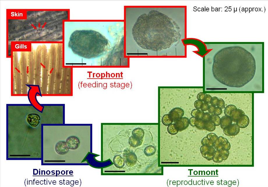
Please can you look at these 4 videos and let me know what Parasite this is ,


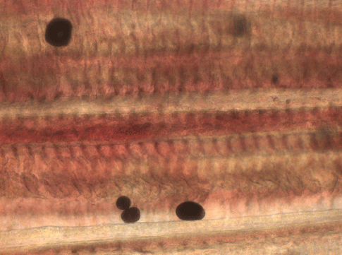
I have also attached a photo .
282950_10200291149382579_1612824924_n.jpg


These were not moving like the above so not sure if relevant.
26-1-17 GOBY 2.jpg


26-1-17 GOBY.jpg
 

Triggreef

Zoa Addict
View Badges
Joined
Apr 11, 2014
Messages
4,929
Reaction score
2,809
Location
East Hampton, CT
Rating - 0%
0   0   0
I don't look under microscope often but i know ich spins like that. I'm pretty sure there are YouTube videos of ich maybe you can compare.
 
OP
OP
Ishy

Ishy

Active Member
View Badges
Joined
May 18, 2016
Messages
136
Reaction score
99
Location
United Kingdom
Rating - 0%
0   0   0
I don't look under microscope often but i know ich spins like that. I'm pretty sure there are YouTube videos of ich maybe you can compare.
Thanks, the ones I've seen are Freshwater but they do seem to look very similar
 
OP
OP
Ishy

Ishy

Active Member
View Badges
Joined
May 18, 2016
Messages
136
Reaction score
99
Location
United Kingdom
Rating - 0%
0   0   0
Thank's, what I don't understand why they arn't all dead they have had this for weeks. The only fish affected are the three tangs and two neon gobies that make contact with them. Thank's for your input.
 

Christoph

Active Member
View Badges
Joined
May 23, 2017
Messages
238
Reaction score
524
Location
Vienna, Austria
Rating - 0%
0   0   0
Hello Ishy,

in my opinion this parasites dont look like amyloodinium ocellatus. - Amyloodinium once feeding look like "pears" attached with one side into the host tissue. This can also be seen well on Humblefishs posted picture (Trophont - feeding stage). The trophonts loose their cilia once attached to the host.

On your videos a lot of cilia can be seen on the surface of the parasite. In my opinion it is cryptocarion irritans. Especially the movement seen in your last video is fairly typical for c. irritans.

Also is amyloodinium way smaller compared to C. irritans. I guess your images were taken at x 400 magnification?

All the best,
Christoph
 
OP
OP
Ishy

Ishy

Active Member
View Badges
Joined
May 18, 2016
Messages
136
Reaction score
99
Location
United Kingdom
Rating - 0%
0   0   0
Thanks for the replies.
I've just uploaded a few more from the same slide if that helps any, quite a few different things seen but we have no idea what we are looking at but we are trying to learn. The microscope is a new bresser 20 -1280 but we are not sure of the magnification on the USB eye piece, we used the 10 x and the 40 x objective.

Please excuse the slide movement on these videos
Thank you for you help


dede

Active Member
View Badges
Joined
Sep 16, 2017
Messages
420
Reaction score
498
Location
North Dakota
Rating - 0%
0   0   0
Interesting, how did you get the skin scrape from your fish, did you use a sedative?
 

Christoph

Active Member
View Badges
Joined
May 23, 2017
Messages
238
Reaction score
524
Location
Vienna, Austria
Rating - 0%
0   0   0
Its certainly very hard to make a diagnosis based only on scrapes, but can be very useful to proof a certain diagnosis based on observation of fish symptoms. The only parasite very easy to identify under the microscope is brooklynella. There are many small critters that dont differ very much from each other.

In the first video from your last post ishy first there can be seen some cryptocarion irritans, but after you change to the 40 x objective there are some "blackish pear shaped dots" that might also be velvet, so you might be looking at a mixed infection, that is imo not very uncommon. Also the size difference seems abour right. So i think @Humblefish is also correct with the initial diagnosis.

Ishy, what macroscopic symptoms/behavioral symptoms do you notice?

Best,
Christoph
 
OP
OP
Ishy

Ishy

Active Member
View Badges
Joined
May 18, 2016
Messages
136
Reaction score
99
Location
United Kingdom
Rating - 0%
0   0   0
Interesting, how did you get the skin scrape from your fish, did you use a sedative?
These were from a Neon Goby no sedative used just held the fish on its side and took the scrape, it took just a couple of seconds. The bigger fish we did before was one person holding the fish and the performing the scrape.
 
OP
OP
Ishy

Ishy

Active Member
View Badges
Joined
May 18, 2016
Messages
136
Reaction score
99
Location
United Kingdom
Rating - 0%
0   0   0
Its certainly very hard to make a diagnosis based only on scrapes, but can be very useful to proof a certain diagnosis based on observation of fish symptoms. The only parasite very easy to identify under the microscope is brooklynella. There are many small critters that dont differ very much from each other.

In the first video from your last post ishy first there can be seen some cryptocarion irritans, but after you change to the 40 x objective there are some "blackish pear shaped dots" that might also be velvet, so you might be looking at a mixed infection, that is imo not very uncommon. Also the size difference seems abour right. So i think @Humblefish is also correct with the initial diagnosis.

Ishy, what macroscopic symptoms/behavioral symptoms do you notice?

Best,
Christoph


IMG_4837[1].jpg

IMG_4835[1].jpg
IMG_4833[1].jpg


Do these help any?

The Lemon peel mimic tang is very pale with the least spots, the spots are difficult to see on the mimic and orange shoulder as they are light coloured fish.
The tangs all display the same symptoms, rubbing on the sand, head twitching, act is something it biting them. For the last 5 days in the morning as lights lights come up they are all in the powerheads turning head on and sideways and generally irritable. By 10 am they calm down, then as lights are almost out the same irritation process but to a lesser degree.
The spots are small not like the usual white spot pictures you see, then there appears to be finer spots covering their bodies, in the eyes of the white cheek and their fins.
All eating
Thanks
 

Christoph

Active Member
View Badges
Joined
May 23, 2017
Messages
238
Reaction score
524
Location
Vienna, Austria
Rating - 0%
0   0   0
Hi,

what i can see on the picture i would interpret as C. irritans, the behavior you mention sounds more like amyloodinium. Probably its a mixed infestation, this is also what the skin scrapes suggest.

All the best,
Christoph
 
OP
OP
Ishy

Ishy

Active Member
View Badges
Joined
May 18, 2016
Messages
136
Reaction score
99
Location
United Kingdom
Rating - 0%
0   0   0
I really appreciate your input on this @Christoph @Humblefish
So given the opinion of mixed infestation would you have an idea why it is only the 3 tangs and 2 gobies ( who clean the tangs) have it and none of the other fish are effected. ?
Also the time scale involved especially with the amyloodinium getting worse over the last few weeks with this week them being in the power heads? Does this not act faster?
Thanks for your time
 

Christoph

Active Member
View Badges
Joined
May 23, 2017
Messages
238
Reaction score
524
Location
Vienna, Austria
Rating - 0%
0   0   0
Amyloodinium can be a very fast killer, but the letality/"virulence" depends on many factors. For example nutrition and the overall state of health of the fish is a key player here. Also some species are more prone to infection than others.

Furthermore if you run a UV-C unit it is likely to keep the amount of infective stages at bay. I have seen tanks where only few fish were affected from a velvet outbreak while in others the outbreak had devastating effect on the whole fish population within a short time frame. I believe that there is differences in "agressiveness" of certain strains of amyloodinium and literature also suggests that fish can obtain a certain amount of immunity against those parasites.

In my opinion you are having two options

a) separating the fish and treating with a medication that is proven effective (chloroquine or copper), letting the display tank run fallow to starve out remaining parasites

b) letting things run its course, support the fish with excellent and frequent food, little stress and a powerful UV-C sanitizer.

I dont want to give you a certain recommendation to choose option A or B, its a heavily debated topic here on R2R. - I guess its best to do a lot of reading and choose the option you feel most comfortable with.

Did you see any fatalities in your fish yet? Do you have the feeling the disease is progressing (getting worse every day)? How is the beathing rate of the fish, do they seem to have troubles getting sufficient oxygen?

All the best, and keep us updated!
Christoph
 

Reefing threads: Do you wear gear from reef brands?

  • I wear reef gear everywhere.

    Votes: 18 14.1%
  • I wear reef gear primarily at fish events and my LFS.

    Votes: 9 7.0%
  • I wear reef gear primarily for water changes and tank maintenance.

    Votes: 1 0.8%
  • I wear reef gear primarily to relax where I live.

    Votes: 19 14.8%
  • I don’t wear gear from reef brands.

    Votes: 72 56.3%
  • Other.

    Votes: 9 7.0%
Back
Top