Clumpy dorsal fins

pureconcept

Community Member
View Badges
Joined
Jan 11, 2021
Messages
73
Reaction score
26
Location
new york city
Rating - 0%
0   0   0
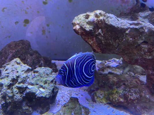
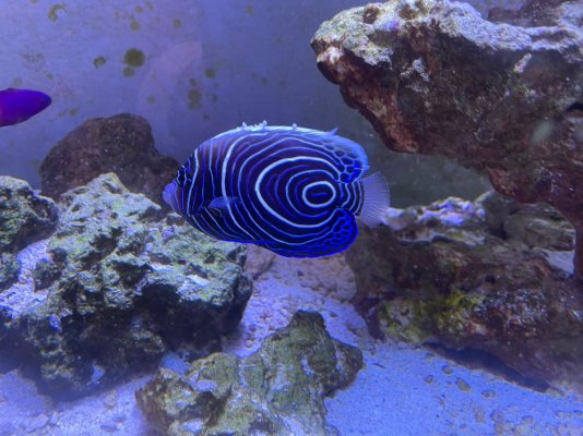
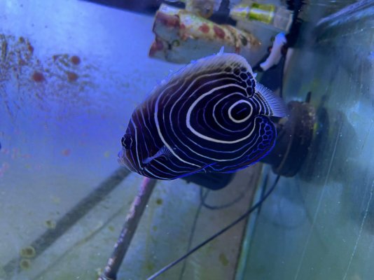
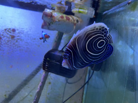
I have an emporer angelfish. It seems to have dots on its fin and body so im taking it out to treat with copper. It also has clumpy dorsal fins. Originally it was just one and i didnt think much of it but now there are multiple clumps on its back. Might the dots be something other than ich and should i be concerned about the fins. Hopefully the pictures are clear enough for someone here to help me out.any help eould be appreciated
IMG-20211021-WA0000.jpg IMG-20211021-WA0002.jpg IMG-20211021-WA0001.jpg IMG-20211021-WA0003.jpg
 

Jay Hemdal

10K Club member
View Badges
Joined
Jul 31, 2020
Messages
26,086
Reaction score
25,846
Location
Dundee, MI
Rating - 0%
0   0   0
I have an emporer angelfish. It seems to have dots on its fin and body so im taking it out to treat with copper. It also has clumpy dorsal fins. Originally it was just one and i didnt think much of it but now there are multiple clumps on its back. Might the dots be something other than ich and should i be concerned about the fins. Hopefully the pictures are clear enough for someone here to help me out.any help eould be appreciated
IMG-20211021-WA0000.jpg IMG-20211021-WA0002.jpg IMG-20211021-WA0001.jpg IMG-20211021-WA0003.jpg


That appears to by Lymphocystis - a viral disease very common to imperator angels. This species is also prone to ich and flukes, but I don't see any clear indication that it has those issues.

How long have you had this fish?
Did you quarantine it?

Here is an excerpt from my upcoming book on fish diseases about Lymphocystis:


Lymphocystis (cauliflower disease)

This is a common viral disease of marine, brackish, and certain freshwater fishes (usually those with marine relatives: glassfish and rainbowfish). It is a chronic (long-lasting) but self-limiting (usually going away on its own) syndrome caused by an iridovirus. The virus causes hypertrophy (enlargement) of the epithelial cells of a fish’s skin and fins.

Initial symptoms consist of off-white to gray nodules on the fish that spread and grow larger over a timeframe of 10 to 90 days. Commonly, when a Lymphocystis nodule forms on a pectoral fin, a new nodule will soon begin to form on the part of the fish that is brushed by the fin, indicating that direct contact can be a mode of spreading the infection.

When Lymphocystis first starts, the small growths can be misdiagnosed as a protozoan infection, such as Cryptocaryon, or even a bacterial infection. The fact that the lesions are long-lasting and do not cause the fish to become acutely ill rules out these more virulent diseases.

Since Lymphocystis is most often seen in newly imported fishes, capture and transport stresses are often mentioned as the stressors that allow this disease to take hold. While this may be true, it may also be that the cause is actually exposure to other infected fish in the aquarium systems of the exporter, importer, or retail suppliers. In any event, it is extremely rare for a fish held in captivity for more than four to six months to suddenly develop this disease.

Treatment with copper sulfate has also been implicated in the development of Lymphocystis in marine fish. The connection is actually not very clear, in that copper sulfate is most often used on newly imported fishes, and those are the ones that develop the disease most often.

Lymphocystis infections can sometimes become more serious, covering large areas of a fish’s body and even interfering with proper feeding if the cell growth involves areas around the mouth. In rare instances, the virus can also cause enlargement of the cells of internal organs, especially in marine fishes (Wolf 1988). This has the potential of causing serious, yet difficult-to-identify diseases in marine fishes.

A variety of cures have been suggested for this disease over the years. Some public aquarists have reported that a reduction in the animal’s environmental stress level will help reduce the severity of an infection. Others have reported that treatment with a mixture of malachite green and formalin (Quick Cure, Formalite, etc.) helps limit the spread of the lesions. Since both of these compounds can damage an aquarium’s bio-filter, and since both are toxic to fish, care must be taken if this method is attempted. By far, the most commonly recommended treatment involves surgically removing the hypertrophied skin cells, followed with a topical antibiotic to hopefully prevent secondary bacterial infection. However, most cases will resolve over time with no treatment needed.

Jay Hemdal
 
OP
OP
P

pureconcept

Community Member
View Badges
Joined
Jan 11, 2021
Messages
73
Reaction score
26
Location
new york city
Rating - 0%
0   0   0
Sorry for late reply but wow ty. That is a very thorough answer. It was quarantined but i didnt think much of the first scale and put it in yhe main tank. As it got worse i put him back i to quarantine. So its a chronic viral infection and i should just give it some time and then let him into the main tank.
 

Tentacled trailblazer in your tank: Have you ever kept a large starfish?

  • I currently have a starfish in my tank.

    Votes: 24 29.6%
  • Not currently, but I have kept a starfish in the past.

    Votes: 18 22.2%
  • I have never kept a starfish, but I hope to in the future.

    Votes: 21 25.9%
  • I have no plans to keep a starfish.

    Votes: 18 22.2%
  • Other.

    Votes: 0 0.0%
Back
Top