Sanity check on current QT process

OP
OP
O

Orion9

Active Member
View Badges
Joined
Jul 9, 2022
Messages
132
Reaction score
58
Location
Missouri
Rating - 100%
2   0   0
I have a microscope now but was concerned about a skin scrape stressing the clown.
The stringy white feces are no longer present in any fish after 3 weeks of metroplex, except for one of my ghost headed blennies who stopped eating two days ago, and was found on its side this morning, with white feces coming out of it. Its in an acclimation box now. These fish were moved to QT on 2/1/23, most are thriving, but now have two dying. I'm confused and would appreciate any guidance or suggestions going forward.
Thanks

 
OP
OP
O

Orion9

Active Member
View Badges
Joined
Jul 9, 2022
Messages
132
Reaction score
58
Location
Missouri
Rating - 100%
2   0   0
About half of the fish are still gill flashing. I had planned a 6hr prazi pro bath at 3x the dosage assuming I still have prazi and hypo salinity resistant flukes. I setup a second QT yesterday and was going to transfer the fish after the bath.
 

Jay Hemdal

10K Club member
View Badges
Joined
Jul 31, 2020
Messages
25,588
Reaction score
25,455
Location
Dundee, MI
Rating - 0%
0   0   0
About half of the fish are still gill flashing. I had planned a 6hr prazi pro bath at 3x the dosage assuming I still have prazi and hypo salinity resistant flukes. I setup a second QT yesterday and was going to transfer the fish after the bath.
Can you look at some of the feces under the scope? That can give you some diagnostics. Skin scrapes take practice, but can show skin parasites. Some people even do gill biopsies, but that requires a fish anesthetic to do safely.
Praziquantel powder can be used as a higher dose dip, but I prefer 2x the normal dose for three hours. I’ve NOT down that with prazipro - I’m worried the glycol solvent it uses could be an issue.

Jay
 
OP
OP
O

Orion9

Active Member
View Badges
Joined
Jul 9, 2022
Messages
132
Reaction score
58
Location
Missouri
Rating - 100%
2   0   0
I have some of the blennies feces on a slide and see a few organisms moving quickly. Any recommendations on resources to match up what I'm seeing with images of parasites.
I believe the blenny has passed and can be used for a skin scrape or gill biopsies.
 
OP
OP
O

Orion9

Active Member
View Badges
Joined
Jul 9, 2022
Messages
132
Reaction score
58
Location
Missouri
Rating - 100%
2   0   0
I'm seeing long, thin, clear worms moving occasionally, and oval shaped, fast moving organisms, that appear to have multiple cells inside, unlike the worms.
 

Jay Hemdal

10K Club member
View Badges
Joined
Jul 31, 2020
Messages
25,588
Reaction score
25,455
Location
Dundee, MI
Rating - 0%
0   0   0
I'm seeing long, thin, clear worms moving occasionally, and oval shaped, fast moving organisms, that appear to have multiple cells inside, unlike the worms.
The biggest issue is knowing if what you are seeing are potential parasites, or are they scavengers that got on the fish after death.
I’ve sometimes been able to take photos through the microscope using my phone - can you try that?
Jay
 
OP
OP
O

Orion9

Active Member
View Badges
Joined
Jul 9, 2022
Messages
132
Reaction score
58
Location
Missouri
Rating - 100%
2   0   0
I was unable to capture pictures and looking for a microscope camera now.
Would scavengers exist in a sterile QT aquarium?
 

Jay Hemdal

10K Club member
View Badges
Joined
Jul 31, 2020
Messages
25,588
Reaction score
25,455
Location
Dundee, MI
Rating - 0%
0   0   0
I was unable to capture pictures and looking for a microscope camera now.
Would scavengers exist in a sterile QT aquarium?
No, not in great numbers.
Jay
 
OP
OP
O

Orion9

Active Member
View Badges
Joined
Jul 9, 2022
Messages
132
Reaction score
58
Location
Missouri
Rating - 100%
2   0   0
I was able to capture the attached video of what I thought looked like a worm, I'm still looking for and trying to capture the fast moving oval bugs. First time using a microscope.
 

Jay Hemdal

10K Club member
View Badges
Joined
Jul 31, 2020
Messages
25,588
Reaction score
25,455
Location
Dundee, MI
Rating - 0%
0   0   0
I was able to capture the attached video of what I thought looked like a worm, I'm still looking for and trying to capture the fast moving oval bugs. First time using a microscope.

That's a nematode worm. Most are non-parasitic, The ones that are parasites, are internal (dog heartworm for example). This one is on a fish scale, so it is external and therefore not parasitic.

That's a nematode worm (round worm). Most are non-parasitic, The ones that are parasites, are internal (dog heartworm for example). This one is on a fish scale, so it is external and therefore not parasitic.

Jay
 
OP
OP
O

Orion9

Active Member
View Badges
Joined
Jul 9, 2022
Messages
132
Reaction score
58
Location
Missouri
Rating - 100%
2   0   0
Would you recommend dissecting the fish and removing part of the gill for observation under microscope? I attempted to extract more feces by putting pressure near the anus but was unsuccessful.
 
OP
OP
O

Orion9

Active Member
View Badges
Joined
Jul 9, 2022
Messages
132
Reaction score
58
Location
Missouri
Rating - 100%
2   0   0
I removed what I believe is part of the gill but did not see anything moving on the slide. The majority of fish are yawning and gill flashing, so I expected to see something on the gill.

IMG-6945.jpg
 

Jay Hemdal

10K Club member
View Badges
Joined
Jul 31, 2020
Messages
25,588
Reaction score
25,455
Location
Dundee, MI
Rating - 0%
0   0   0
Gills are always something to look at, but that needs to be done with a fish that’s been dead less than 30 minutes or so.
You want to see dark red gills, no moving parasites and no missing or eroded gill tissue.
Jay

Would you recommend dissecting the fish and removing part of the gill for observation under microscope? I attempted to extract more feces by putting pressure near the anus but was unsuccessful.
 

Jay Hemdal

10K Club member
View Badges
Joined
Jul 31, 2020
Messages
25,588
Reaction score
25,455
Location
Dundee, MI
Rating - 0%
0   0   0
That seems too high of magnification.
Jay
 
OP
OP
O

Orion9

Active Member
View Badges
Joined
Jul 9, 2022
Messages
132
Reaction score
58
Location
Missouri
Rating - 100%
2   0   0
The gill was examined 6-10 hours after the fish passed, so I missed the window of opportunity. I studied the slides at the lowest magnification first and kept increasing hoping to see something moving. Am I looking for organisms that are similar in size the the roundworm I captured, or larger?
 

Jay Hemdal

10K Club member
View Badges
Joined
Jul 31, 2020
Messages
25,588
Reaction score
25,455
Location
Dundee, MI
Rating - 0%
0   0   0
The gill was examined 6-10 hours after the fish passed, so I missed the window of opportunity. I studied the slides at the lowest magnification first and kept increasing hoping to see something moving. Am I looking for organisms that are similar in size the the roundworm I captured, or larger?
Trematodes will be about the same size or slightly smaller than the roundworm. Protozoans will be about as big as the worm’s width to perhaps 2x its width.
Jay
 
OP
OP
O

Orion9

Active Member
View Badges
Joined
Jul 9, 2022
Messages
132
Reaction score
58
Location
Missouri
Rating - 100%
2   0   0
While examining feces yesterday I saw several fast moving oval shaped protozoans that came to point at one end. They were slightly wider than the roundworm but not very long, about twice its own width.

As of today they've been in hypo salinity for 5 weeks and metroplex dosed every 48hrs for 3 weeks. Previously treated with Copper Power, metroplex, General Cure, and prazi beginning in November 2022 while they were still in the DT, copper was after DT.

I'm considering the 2x strength prazi pro bath for 3 hours while in the current QT, then change 50% water after 2hrs, repeat in 5-7 days, then move to a sterile second QT that has been cycling for a few days.

Does this sound like a logical approach? I believe flukes are still present due to constant gill flashing and yawning.
 

Jay Hemdal

10K Club member
View Badges
Joined
Jul 31, 2020
Messages
25,588
Reaction score
25,455
Location
Dundee, MI
Rating - 0%
0   0   0
Generally, (but not always) fast moving protozoans are not parasitic.

Your plan sounds fine for flukes, I just wish we had a positive diagnosis for that.

Jay
 
OP
OP
O

Orion9

Active Member
View Badges
Joined
Jul 9, 2022
Messages
132
Reaction score
58
Location
Missouri
Rating - 100%
2   0   0
Prazi bath is complete. all fish are eating and acting normal.

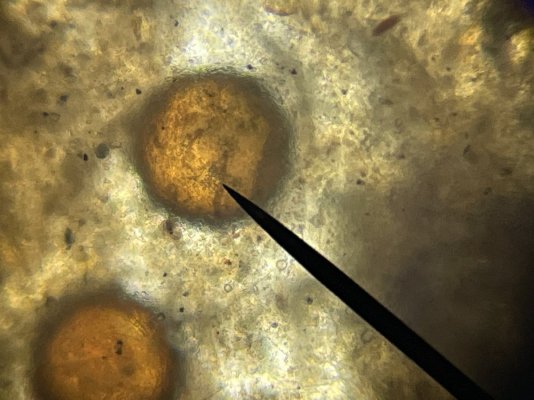
My remaining ghost blenny had a long sting of brownish white feces I was able to capture and review. Nothing was moving but I found these images interesting, almost looks like hatched and unhatched eggs?


IMG-6957.jpg

IMG-6948.jpg

IMG-6959.jpg
 

Attachments

  • IMG-6948.jpg
    IMG-6948.jpg
    182 KB · Views: 16

More than just hot air: Is there a Pufferfish in your aquarium?

  • There is currently a pufferfish in my aquarium.

    Votes: 30 17.2%
  • There is not currently a pufferfish in my aquarium, but I have kept one in the past.

    Votes: 29 16.7%
  • There has never been a pufferfish in my aquarium, but I plan to keep one in the future.

    Votes: 32 18.4%
  • I have no plans to keep a pufferfish in my aquarium.

    Votes: 75 43.1%
  • Other.

    Votes: 8 4.6%
Back
Top