
OP
OP
taricha
5000 Club Member
View Badges
Partner Member 2024
Excellence Award
Article Contributor
R2R Research
- Joined
- May 22, 2016
- Messages
- 6,575
- Reaction score
- 10,162
That's an awesome paper for this discussion. Thank you!Is it possible to roughly translate pigment composition percentages to action spectra? When you look at the pigment concentrations in this study. It would appear that the action spectra would be very different between the acro and brain corals.
Also, yes. I think since it gives us pigment mass concentrations, we can build an absorption spectrum for the zoox. And as the last few posts discussed - it looks a lot like an action spectrum (give or take a few quirks).
I'll give it a shot anyway. See if the result is sensible.
P.s. and a tidbit for later...
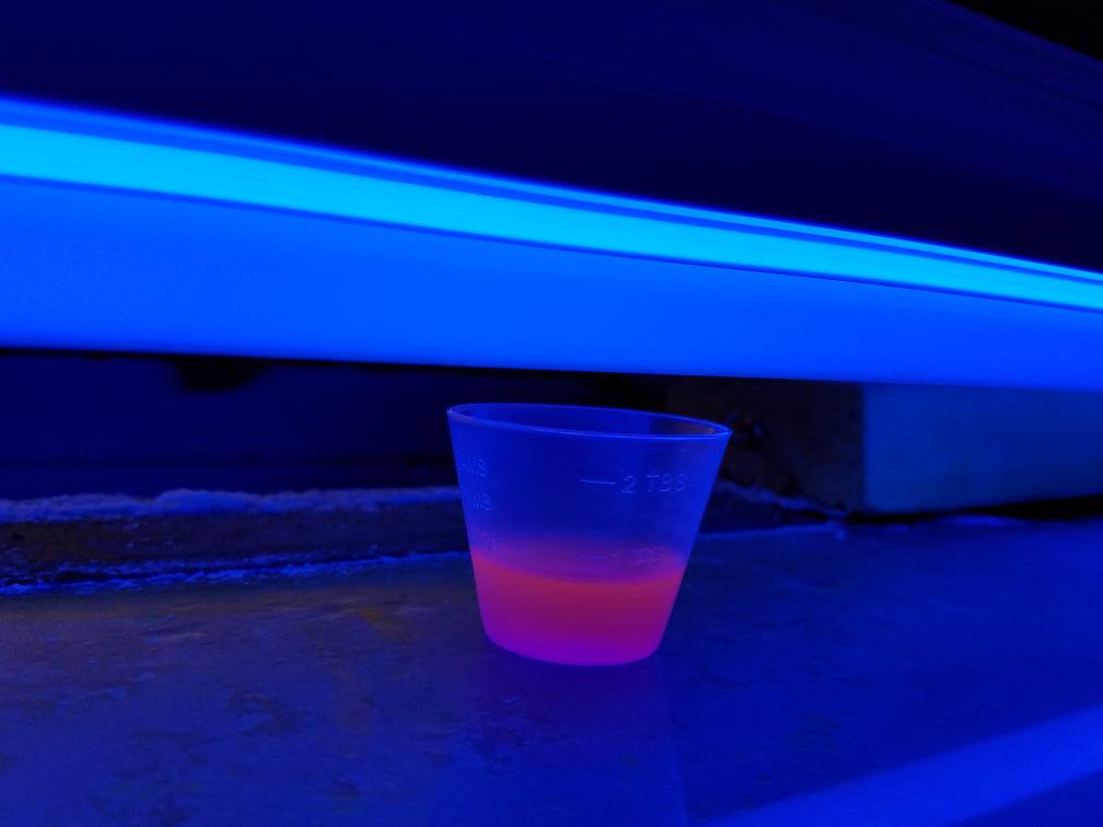
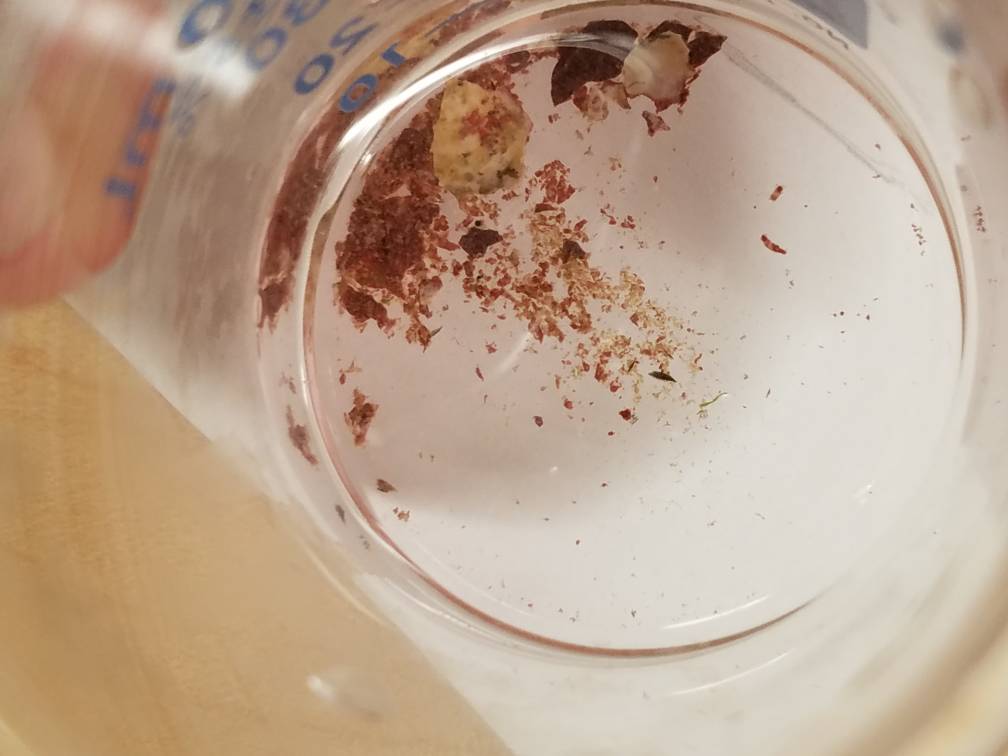
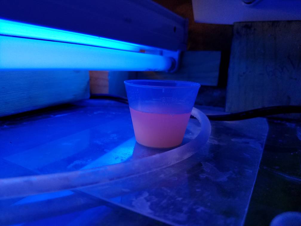
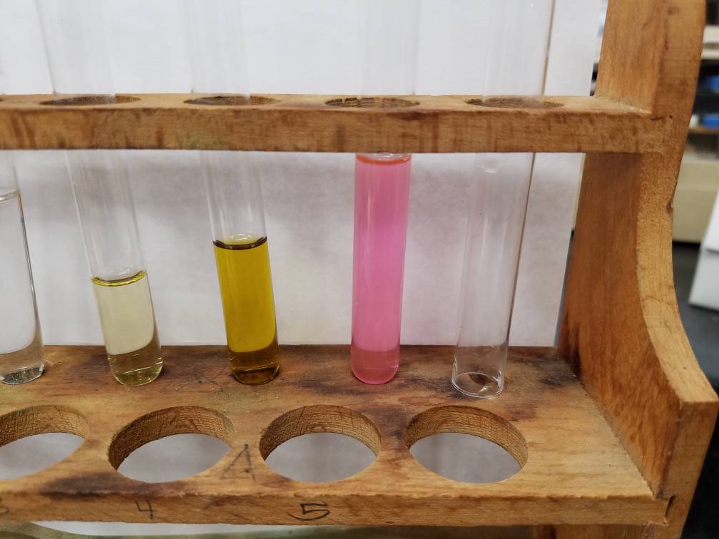
What photosynthetic organism in my tank has this insane pink as its pigments? [emoji848]
(All that was done to it was freeze, smash, thaw, pour through paper towel)
Last edited: