Not my thread, but It seems pretty perfectly on-topic so.....
Brown Jelly Disease in 1 day melted some purple mushrooms that have made it through a decade of me largely not knowing what I was doing, and survived multiple tank disasters.
Nothing else in the tank affected (sps, LPS, NPS, softies, gorgs, nems etc etc.) Zero chance these were "stressed" when nothing else in the tank is. Zero chance that they were dying on their own and brown jelly moved in. anyway.
pics yesterday. The healthy purple mushroom on the left in the 2nd pic has since been overtaken by the blob.
as many people elsewhere have reported and as I have seen in my tank before when brown jelly ate some xenias (btw, convenient it attacks corals I care nothing about. lucky me) - Brown Jelly is marked by an overwhelming swarm of ciliates.
These ciliates have ingested the zooxanthellae (brown dots) from the coral tissue.
playlist with two videos: first low power shows the raft of ciliates forming the brown jelly structure. 2nd higher power shows Ciliates having ingested zooxanthellae, ciliates burrowing into matter to feed, tiny bacteria-scale zooming dots, and unknown (ID help?) motionless jellybean shapes with curled lines inside.
If the hobby knowledge on this disease has progressed past this, then please share.
I have metroplex (metronidazole) on hand, and chemi-clean (erythromycin). I started a treatment of the metro today (2 scoops per 10gal) to see the effects on the ciliates, the bacteria, and the overall brown goo and coral.
I'll test the erythromycin after I'm convinced of the outcome of the metro.
Over my head but that last pdf was pretty interesting - I had "something" go thru my tank & it seemed bacteria dosing made it back off/go away. That was acros to a goniopora & then torch then back to acros tho, would spread once it had killed what it was on last.
Today, added second metro dose - 24hr post metro 1st dose, no noticeable change in the Brown jelly. Bacteria, zooxanthellae, ciliates - all seem unchanged. It almost fully dissolved the handful of affected purple mushrooms.
It also hopped to a nearby Kenya tree stalk, and a green mushroom on opposite side of the tank.
Perhaps my imagination, but under the scope there seemed to be more bacterial activity at the site of new infections and much less visible bacterial wiggles at the older blobs.
I think if no change after 48hr, I'll see what chemiclean does.
I would try API EM erythromycin, unless you've solved the mystery of what form of erythromycin Chemiclean contains.
I only know it does not contain erythromycin succinate.
I would try API EM erythromycin, unless you've solved the mystery of what form of erythromycin Chemiclean contains.
I only know it does not contain erythromycin succinate.
I think your roving pack of hungry ciliates might be paramecium.
The single round objects are probably cocci, type unknown.
Remember macrolides (erythromycin) are a time dependent antimicrobial assuming your trial dose meets MIC for the intended subject.
So don't judge your results in less than about 72 hours I'd think.
Are you conducting your kill studies in a dish or on a growth plate?
You need to make sure the little buggers have food to sustain themselves while you try to nuke 'em.
From the original posters? Nothing that I know of, but someone feel free to point us to something otherwise.
I'll update as long as I have an infection to test on, and treatments to try.
ID follow-up the tiny jelly beans with curls inside are nematocysts from the coral!
Remember macrolides (erythromycin) are a time dependent antimicrobial assuming your trial dose meets MIC for the intended subject.
So don't judge your results in less than about 72 hours I'd think.
Are you conducting your kill studies in a dish or on a growth plate?
I take your point about the time, but given the spread rate of BJD, something that doesn't slow it down until 72hr is not super helpful anyway.
The metro test has been in-tank.
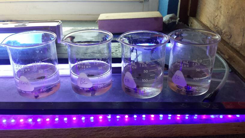
But today, I took healthy cuttings of a green mushroom, purple mushroom, and Kenya tree (3 types that have been affected in this outbreak) put pieces in 4 beakers with 100ml tank water.
I sucked up a couple blobs of active brown jelly infection, mixed it up and added equal parts to each beaker.
Then one beaker got chemiclean, one the e.m. Erythromycin from api, one got api's doxycycline, and control got nothing. Each was at double the label recommended concentration.
I also found a 2014 paper that seems to advance the investigation a good bit. I'll post it and key takeaways later, but the upshot is that they spread the infection to healthy corals using sub-micron filtered material, and halted the infection with some antibiotics but not others, strongly indicating a bacterial cause.
Vibrio bacteria got blamed early, but researchers found no difference in the amounts of vibrio in healthy and diseased coral.
Doesn't mean it's not bacteria, though.
Well, stopping the spread of a bacterial infection vs eradication of the colony to the point it can't recover can be very different.
I applaud your efforts to just accomplish the first step which would be to identify the causative organism, if there actually is one.
Finding the correct agent for MIC and minimum contact time for bactericidal effect can take some dedication and time.
If you are successful you will have done the reefing community a great service.
Many blessings upon your efforts.
... WBD [White Band Disease] is amenable to treatment with either ampicillin or paromomycin sulfate
WBD is, in fact, a transmissible disease attributable to a 0.22–0.45 µm filterable fraction that was susceptible to antibiotic treatment. (likely Gram Positive Bacteria)
we conclude that WBD is caused by microorganisms rather than by physiological stress.
This study also shows that WBD, and other diseases such as WS [White Syndrome], white plague, and black band disease is a polymicrobial disease associated with a number of specific microorganisms that are consistently associated with diseased samples but absent, or undetectable, in healthy ones.
One of these, the ciliate Philaster lucinda (KC832299), has recently been shown to be consistently associated with the coral disease WS in the Pacific [17] and within aquaria [28], which all have identical disease signs. (this is the ciliate that is characteristic of Brown Jelly)
However, selective elimination of [ciliate] using metronidazole failed to arrest disease lesion progression in controlled experiments...
However, ...analysis showed that the ...disease changed in the metronidazole treatment. This result is consistent with this ciliate being a secondary pathogen which nonetheless contributes to the typical pathogenesis of WBD and WS. (The ciliate's presence/absence determines the look of the disease.)
Is a combination of more than one of these three potential primary bacterial pathogens required to maintain the disease state, although all three plus the ciliate consistently co-occurred in the disease in nature?
Ok. This is not going as expected. Which I guess is what makes it an experiment. :)
tagging @jason2459
The in-tank treatment of two doses of metroplex (metronidazole) over 2 days at 2x the label dosage has now gone 72 hr.
Brown jelly continued to slowly spread and eat a mushroom or two here and there.
Inspection of the brown jelly itself shows no noticeable changes in any of the components of the jelly. Bacteria, Ciliates, and Zooxanthellae are all still present in great quantities and seem unaffected. Contrary to the paper I cited above "Treatment with metronidazole reduced [the ciliate] to below detection limits, but did not arrest the disease. However, the microscopic disease signs changed, suggesting a secondary role in disease causation for this ciliate." I'll have to dig in and see what doses they were using.
Regardless, Metro at recommended doses did nothing.
On to the 4 beakers: Control (nothing), Chemiclean, E.M. Erythromycin(API), and API Fin and Body Cure (Doxycycline hyclate)
After 24 hours.
By visual inspection, the water:
Control: cloudy with tons of ciliates
Chemiclean (CC): milky with ciliates and bacteria
Erythromycin (EM): clear water
Doxycycline (DC): clear pink water (the label warns about water discoloration)
The corals in each beaker fared about the same in the control, CC, and EM beakers: the green mushroom, purple mushroom, and kenya tree all became covered in brown jelly overnight in each of those 3 beakers. In fact, in the control the purple mushroom piece had been dissolved completely.
Inspection of the Brown Jelly itself in all 3 beakers showed the same thing - no change - lots of ciliates, lots of bacterial wiggles. I have no way of telling if some classes of bacteria had been knocked out by the EM or CC vs the control.
Except the Doxycycline (DC).
In the DC, there was very little brown jelly, and no signs of the coral pieces added getting overrun by infection.
For comparison...
first, Control - cloudy with masses of ciliates
2nd CC
3rd EM
4th DC
I was amazed at how clean and almost totally jelly-free the cuts in the Doxycycline were, I put it under a scope to look closer.
super clean - no noticeable tissue degradation at the cut site. almost totally Brown Jelly free.
Examining the small amounts of brown jelly that were in the DC beaker under the scope, there were still tons of ciliates, but almost no visible bacterial wiggles.
See the microscope video for comparison of the almost absent bacterial wiggles - first in the DC.
now compare that to the large amount of visible bacterial activity in the CC
This is all very interesting, however the label on the box says "for use in Freshwater aquariums," so without further testing or information, I'd have to say putting this in a reef tank would be reckless and stupid. I can't imagine that a reef tank biofilter would do okay.
However, I did poke around and found a few other people also saying doxycycline is a usable treatment for a coral colony outside a tank. It sure looks promising after 24 hours.
Hm.....I don't know anything specifically about BJD but the articles and ideas posted are leading me to think about carbon loading in the Caribbean as a precursor to anything going on there.
Carbon loading tweaks the whole microbial scene away from corals and phytoplankton communities.
I'm also led to think about an article I read that observed that pathogenic relationships are "turned up" in virulence when in a disturbed environment.
I'm not aware of anything Earthly that stresses or otherwise harms mushrooms but the specificity of the breakout in @taricha's tank does seem to indicate that the shrooms were particularly stressed and/or susceptible for some reason compared to neighboring colonies which did not succumb to BJD.
It may seem odd for one coral to be the only target (still does to me), but I recently had an issue that was precisely long enough to kill about 1.2 square feet of Montipora digitata colony across 3-4 light zones and about as many different flow zones. At the same time, no other corals in the whole system appeared to suffer at all. I can't explain the specificity in either case except to presume some kind of stress.
All just 2¢. Very interesting topic!!!
@taricha any idea when/where/why/how your BJD got started?
And BTW, I might be willing to pay for shipping on some BJD for my tank if you can prove this stuff will kill my mushrooms off once and for all!!! ;) ;) ;)
I can't come up with any "stress" that these mushrooms might be experiencing. (nor any recent addition that could have brought a new pathogen)
I mean over the years these mushrooms have survived being dropped in a plastic container from 2nd story balcony onto concrete, brush scrubbed, cut many times, blasted with flow until they detached, stinging coral warfare, 95+degree overheating, covered in kalk paste, aptasiaX, left out of water for a couple hours, all kinds of medication/oxidizer/algaecide tests, violent swings in salinity and every other water parameter, I even injected a mL or 2 of peroxide directly into gut chambers a couple of times.
None of those things allowed an infection of the mushrooms. At best they hurt a small section of tissue, and the rest grew back happily. And right now, all is well in the tank. nothing else in the tank is unhappy. It's hard to point to "stress" as a cause here while keeping a straight face.
Seems like a strong pathogenic effect is a much bigger driver here than stress. That's why I decided to go pathogen hunting rather than chasing down some hidden water quality boogey man.
follow-ups. after 2 days in the beakers, everything was dead. I suppose no aeration in stagnant beakers was a problem.
Took more healthy mushroom cuttings, and put them in an aerated beaker along with brown jelly. I'll see if I can start the infection, and then halt the infection in progress with doxycycline, and save the infected pieces.
The likely reason I couldn't reproduce the study's results with metronidazole wiping out the ciliates was that the study used 100ppm of metro. The recommended in-tank dose is 2.5 ppm, and I doubled it to 5ppm.
Though metro is used as a tank-wide treatment, at 100ppm, it'd kill a LOT of stuff. I'll probably try it in a beaker, just to see what BJD without the ciliates looks like.