Mycobacterium? Something else?

kels64

Community Member
View Badges
Joined
Mar 8, 2020
Messages
81
Reaction score
86
Rating - 0%
0   0   0
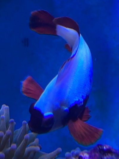
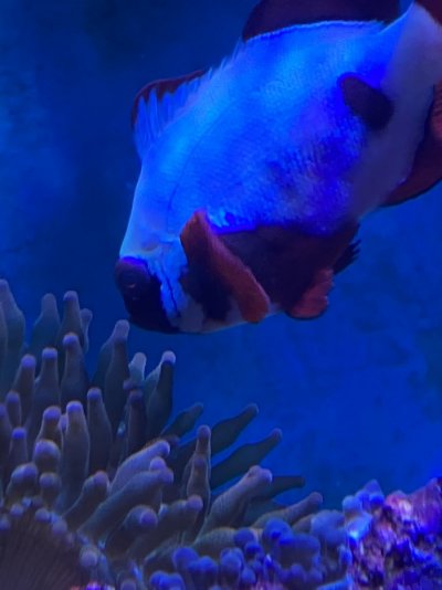
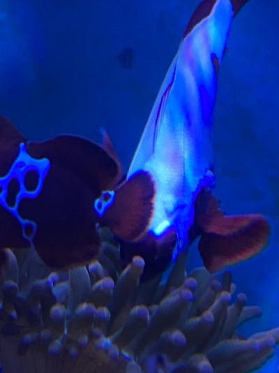
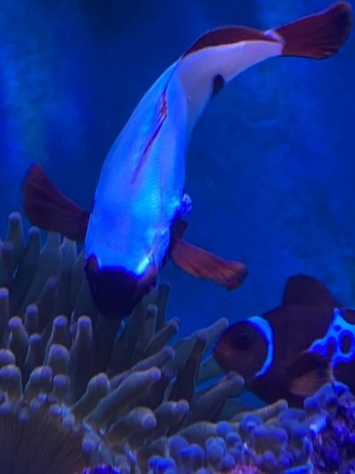
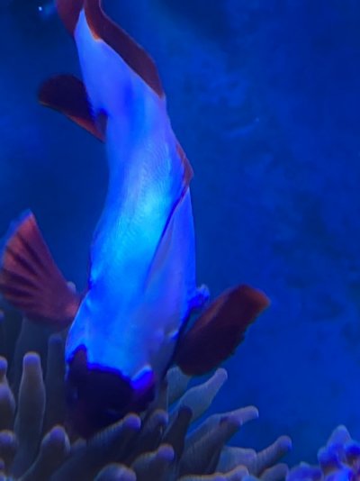
So I’ve had this fish for about a month and she has been a head down swimmer ever since I got her so we all know clown fish are weird and I didn’t think anything of it. Today I turned up the lights to do my weekly close check of everybody and see that she has some kind of lesion/tumor and also appears to have a Popeye on one side. I’m including video if anybody can give me clarification of what they think this looks like. I’m concerned about tuberculosis obviously. The male lightning maroon that is in the tank with her fine. He has no spots or lesions or symptoms. She is not fully mature, less than a year old. Gold flake maroon. Of course this is my favorite fish in my favorite anemone tank. My heart is broken unless you guys can recommend some kind of process or treatment and maybe it’s not what I fear it is. C5F6A14E-4D6E-42B2-85BC-81314ED0C309.jpeg 0CC4EA52-42F0-4EEA-8C03-CB37346F214A.jpeg 130241A5-C647-44A0-B524-9B35D773443F.jpeg 31169323-8582-4AB2-97D5-93E173572C84.jpeg 5D921013-C9C0-4C5A-A0A8-35AA34D0A3D5.jpeg
 

Attachments

  • IMG_3949.MOV
    80.2 MB

vetteguy53081

Well known Member and monster tank lover
View Badges
Joined
Aug 11, 2013
Messages
91,695
Reaction score
202,386
Location
Wisconsin -
Rating - 100%
13   0   0
It doesnt appear to be fungal but bacterial and even with bacterial closer to Lymphocystis. ymphocystis appears as a white or beige colored wart-like growth that usually starts on the fins and spines and sometimes spreads to the body. It can also grow over internal organs, including a fish’s gills. Initially it may be small (looks like Ich), but then grows in size (which is how you know it’s not Ich). Lympho is a virus that many fish carry for life.
So How did I come up with this thought?
The most obvious sign is the appearance of small to moderate-sized, irregular, nodular, wart-like growth which appears in video. . A strong tentative diagnosis of lymphocystis can be made based on seeing round to oval, grape, or balloon like structures, often in clumps. For an accurate diagnosis, a sample under a microscope would help determine.
Lymphocystis nodules may also be confused with epitheliocystis (an intracellular bacterial disease), clumps of fungus, or mucus tags. Some skin neoplasias (cancers) may also be confused with lymphocystis. Because of the potential confusion with other disease agents, diagnosis of lymphocystis should be verified.
If a fish has numerous lesions and/or lymphocystis nodules cover a large portion of important organs (e.g., the gills), there may be some impairment and greater chance of secondary infections by bacteria, parasites, or fungi that can now more easily infect the fish and help contribute to mortalities. Maracyn 2 comes to mind as a possible treatment.
A person who works with diseases is @Jay Hemdal
Lets see what he thinks on this also.
 
OP
OP
kels64

kels64

Community Member
View Badges
Joined
Mar 8, 2020
Messages
81
Reaction score
86
Rating - 0%
0   0   0
It doesnt appear to be fungal but bacterial and even with bacterial closer to Lymphocystis. ymphocystis appears as a white or beige colored wart-like growth that usually starts on the fins and spines and sometimes spreads to the body. It can also grow over internal organs, including a fish’s gills. Initially it may be small (looks like Ich), but then grows in size (which is how you know it’s not Ich). Lympho is a virus that many fish carry for life.
So How did I come up with this thought?
The most obvious sign is the appearance of small to moderate-sized, irregular, nodular, wart-like growth which appears in video. . A strong tentative diagnosis of lymphocystis can be made based on seeing round to oval, grape, or balloon like structures, often in clumps. For an accurate diagnosis, a sample under a microscope would help determine.
Lymphocystis nodules may also be confused with epitheliocystis (an intracellular bacterial disease), clumps of fungus, or mucus tags. Some skin neoplasias (cancers) may also be confused with lymphocystis. Because of the potential confusion with other disease agents, diagnosis of lymphocystis should be verified.
If a fish has numerous lesions and/or lymphocystis nodules cover a large portion of important organs (e.g., the gills), there may be some impairment and greater chance of secondary infections by bacteria, parasites, or fungi that can now more easily infect the fish and help contribute to mortalities. Maracyn 2 comes to mind as a possible treatment.
A person who works with diseases is @Jay Hemdal
Lets see what he thinks on this also.
Thank you thank you thank you so much! I am relieved to hear that you don’t really think it looks like mycobacterium. Can you give me information on what long-term prognosis may be for this fish? And what you mentioned the Maracyn 2, is that an antibiotic treatment? Can you tell me if this condition is contagious? There’s only one other fish in there with her which is her mate.
 
OP
OP
kels64

kels64

Community Member
View Badges
Joined
Mar 8, 2020
Messages
81
Reaction score
86
Rating - 0%
0   0   0
It doesnt appear to be fungal but bacterial and even with bacterial closer to Lymphocystis. ymphocystis appears as a white or beige colored wart-like growth that usually starts on the fins and spines and sometimes spreads to the body. It can also grow over internal organs, including a fish’s gills. Initially it may be small (looks like Ich), but then grows in size (which is how you know it’s not Ich). Lympho is a virus that many fish carry for life.
So How did I come up with this thought?
The most obvious sign is the appearance of small to moderate-sized, irregular, nodular, wart-like growth which appears in video. . A strong tentative diagnosis of lymphocystis can be made based on seeing round to oval, grape, or balloon like structures, often in clumps. For an accurate diagnosis, a sample under a microscope would help determine.
Lymphocystis nodules may also be confused with epitheliocystis (an intracellular bacterial disease), clumps of fungus, or mucus tags. Some skin neoplasias (cancers) may also be confused with lymphocystis. Because of the potential confusion with other disease agents, diagnosis of lymphocystis should be verified.
If a fish has numerous lesions and/or lymphocystis nodules cover a large portion of important organs (e.g., the gills), there may be some impairment and greater chance of secondary infections by bacteria, parasites, or fungi that can now more easily infect the fish and help contribute to mortalities. Maracyn 2 comes to mind as a possible treatment.
A person who works with diseases is @Jay Hemdal
Lets see what he thinks on this also.
Also, I’m wondering if you can refer me to somewhere where I can get microscopic images of this? I am a laboratory professional and I do have a microscope here at home and I would be happy to clip a little tissue off and look at it if I know what I’m looking for.
 

vetteguy53081

Well known Member and monster tank lover
View Badges
Joined
Aug 11, 2013
Messages
91,695
Reaction score
202,386
Location
Wisconsin -
Rating - 100%
13   0   0
Also, I’m wondering if you can refer me to somewhere where I can get microscopic images of this? I am a laboratory professional and I do have a microscope here at home and I would be happy to clip a little tissue off and look at it if I know what I’m looking for.
In my area, would be a college or college extension.
Ive never done this myself but bet @Jay Hemdal has
 

Jay Hemdal

10K Club member
View Badges
Joined
Jul 31, 2020
Messages
25,677
Reaction score
25,529
Location
Dundee, MI
Rating - 0%
0   0   0
I generally try to avoid diagnosing things as Mycobacteriosis, as it is an easy trap to fall into - "I see mixed vague symptoms, so it must be Myco". The reality is that Mycobacterium marinum, and to a lesser extend, two other species, chelonae and fortuitum are more or less found in every aquarium environment - even in the food we feed out fish. However, fish can harbor chronic infections for years, but generally do not succumb to acute infections until they are old.

The exophthalmia and the lesion may be Myco, but I doubt it. They may not even be directly related to one another. The eye may resolve on its onw, the lesion is what worries me - it doesn't look like typical Lymphocystis to me - wrong location, and darker in color and more deeply "rooted" than I would expect Lymphocystis to be. However, there are other viruses that can cause growths like this. I've seen them on walleye, but never a clownfish.

How long has it had this growth?

Jay
 
OP
OP
kels64

kels64

Community Member
View Badges
Joined
Mar 8, 2020
Messages
81
Reaction score
86
Rating - 0%
0   0   0
I generally try to avoid diagnosing things as Mycobacteriosis, as it is an easy trap to fall into - "I see mixed vague symptoms, so it must be Myco". The reality is that Mycobacterium marinum, and to a lesser extend, two other species, chelonae and fortuitum are more or less found in every aquarium environment - even in the food we feed out fish. However, fish can harbor chronic infections for years, but generally do not succumb to acute infections until they are old.

The exophthalmia and the lesion may be Myco, but I doubt it. They may not even be directly related to one another. The eye may resolve on its onw, the lesion is what worries me - it doesn't look like typical Lymphocystis to me - wrong location, and darker in color and more deeply "rooted" than I would expect Lymphocystis to be. However, there are other viruses that can cause growths like this. I've seen them on walleye, but never a clownfish.

How long has it had this growth?

Jay
Jay thank you so much for your response. This happened literally within the last couple of days. Although you can’t really tell from the fuzzy pictures and probably the videos not great either but it actually tends to look like it has a little stalk to it. She has had the appearance of what I would call elevation of the skin area in a certain spot which you can still see in the pictures and video but the lesion/tumor is brand new. I look at them several times a day and weekly I do a all lights on pretty thorough check and this is not anything that had been on her on Thursday. I know that because on Thursday I walked out and looked at the tank and she was laying on top of the spray bar at the surface of the water. I thought she was dead. I picked her up and looked at her and I didn’t see any marks on her and then she twitched her tail so I placed my hand down in the water and she swam out of my hand and into her anemone. She had not had that behavior before and she has not displayed it since. In all other regards she seems to be fine, hosting well with her anemone and interacting with her mate and eating.
 

Jay Hemdal

10K Club member
View Badges
Joined
Jul 31, 2020
Messages
25,677
Reaction score
25,529
Location
Dundee, MI
Rating - 0%
0   0   0
If you had some fish anesthetic, I would suggest snipping it off, but without that it would be too risky. I guess just watch it for now?
Jay
 
OP
OP
kels64

kels64

Community Member
View Badges
Joined
Mar 8, 2020
Messages
81
Reaction score
86
Rating - 0%
0   0   0
I’m comfortable with that plan. I will just keep an eye on her and watch for any change in behavior, eating, or mobility. Thank y’all so much for your advice and for helping me (and her). I feel more optimistic about her condition after hearing what you guys had to say. I really appreciate you!
 
OP
OP
kels64

kels64

Community Member
View Badges
Joined
Mar 8, 2020
Messages
81
Reaction score
86
Rating - 0%
0   0   0
Sadly my big girl died this morning. Upon examination I found several ulcers that had developed overnight on her side as well as and extrusion of white material from her cloacal opening. Upon microscopic examination I was unable to determine what this was but it did not appear to be parasite. It almost had the appearance of a fatty material. I am deeply saddened and of course now concerned for her mate. At this time he is apparently very healthy and seems to have no blemishes nor disease process.
My heart is broken.
 

Jay Hemdal

10K Club member
View Badges
Joined
Jul 31, 2020
Messages
25,677
Reaction score
25,529
Location
Dundee, MI
Rating - 0%
0   0   0
The white material was likely mucus or sloughed cells from the intestinal lining, not sure what that means though when combined with external lesions, other than it was likely a systemic issue- sorry!
Jay
 
OP
OP
kels64

kels64

Community Member
View Badges
Joined
Mar 8, 2020
Messages
81
Reaction score
86
Rating - 0%
0   0   0
The white material was likely mucus or sloughed cells from the intestinal lining, not sure what that means though when combined with external lesions, other than it was likely a systemic issue- sorry!
Jay
Thanks Jay I appreciate that. It makes me very sad and her mate looks very lonely.
 

RobertTheNurse

Well-Known Member
View Badges
Joined
May 3, 2020
Messages
855
Reaction score
707
Rating - 0%
0   0   0
It doesnt appear to be fungal but bacterial and even with bacterial closer to Lymphocystis. ymphocystis appears as a white or beige colored wart-like growth that usually starts on the fins and spines and sometimes spreads to the body. It can also grow over internal organs, including a fish’s gills. Initially it may be small (looks like Ich), but then grows in size (which is how you know it’s not Ich). Lympho is a virus that many fish carry for life.
So How did I come up with this thought?
The most obvious sign is the appearance of small to moderate-sized, irregular, nodular, wart-like growth which appears in video. . A strong tentative diagnosis of lymphocystis can be made based on seeing round to oval, grape, or balloon like structures, often in clumps. For an accurate diagnosis, a sample under a microscope would help determine.
Lymphocystis nodules may also be confused with epitheliocystis (an intracellular bacterial disease), clumps of fungus, or mucus tags. Some skin neoplasias (cancers) may also be confused with lymphocystis. Because of the potential confusion with other disease agents, diagnosis of lymphocystis should be verified.
If a fish has numerous lesions and/or lymphocystis nodules cover a large portion of important organs (e.g., the gills), there may be some impairment and greater chance of secondary infections by bacteria, parasites, or fungi that can now more easily infect the fish and help contribute to mortalities. Maracyn 2 comes to mind as a possible treatment.
A person who works with diseases is @Jay Hemdal
Lets see what he thinks on this also.
I just love when people speak all medical lol very cool
 

vetteguy53081

Well known Member and monster tank lover
View Badges
Joined
Aug 11, 2013
Messages
91,695
Reaction score
202,386
Location
Wisconsin -
Rating - 100%
13   0   0

Courtney Aldrich

Active Member
View Badges
Joined
Dec 31, 2020
Messages
212
Reaction score
266
Location
Minneapolis
Rating - 0%
0   0   0
Can you send some of the tissue to a microbiology lab to see if they can culture the pathogen? If you have M. marinum in your tank you should be careful and make sure to wear gloves. Non-tuberculous mycobacterial (NTM) infections can be very challenging to treat (see this information at National Jewish Health, which is one of the premiere clinics in the US for treating NTMs: https://www.nationaljewish.org/cond...bacteria-overview/types/mycobacterium-marinum). While M. marinum may be common in fresh and saltwater, I can't believe it is found in all aquariums. Aquabiomics has done microbiome testing and has a panel for common pathogens including mycobacteria. I haven't seen anyone post their results with M. marinum.
 
Last edited:
OP
OP
kels64

kels64

Community Member
View Badges
Joined
Mar 8, 2020
Messages
81
Reaction score
86
Rating - 0%
0   0   0
Can you send some of the tissue to a microbiology lab to see if they can culture the pathogen? If you have M. marinum in your tank you should be careful and make sure to wear gloves. Non-tuberculous mycobacterial (NTM) infections can be very challenging to treat (see this information at National Jewish Health, which is one of the premiere clinics in the US for treating NTMs: https://www.nationaljewish.org/cond...bacteria-overview/types/mycobacterium-marinum). While M. marinum may be common in fresh and saltwater, I can't believe it is found in all aquariums. Aquabiomics has done microbiome testing and has a panel for common pathogens including mycobacteria. I haven't seen anyone post their results with M. marinum.
Thank you for the information Courtney I’m not convinced that that’s what it is/was. The other fish is perfectly healthy and I guess in time we will see. I’m beginning to wonder if it wasn’t a genetic issue that was contributing rather than just a disease process. There seemed to be a myriad of issues that would come and go. Anyway, her body has been disposed of and hopefully there will be no further problems. I plan to keep her mate alone for a while to observe for any indications of illness. If a month passes with no indication of any problems I will think about getting him a new mate. Being a maroon, this will be a challenge. Hopefully I will be able to find a HEALTHY gold nugget like the beautiful girl I lost.
 

Being sticky and staying connected: Have you used any reef-safe glue?

  • I have used reef safe glue.

    Votes: 141 88.1%
  • I haven’t used reef safe glue, but plan to in the future.

    Votes: 9 5.6%
  • I have no interest in using reef safe glue.

    Votes: 7 4.4%
  • Other.

    Votes: 3 1.9%
Back
Top