- Joined
- Apr 22, 2020
- Messages
- 28
- Reaction score
- 9
Hi everyone,
I am a tank caretaker at our college and we have this interesting case I wanted to share in case anyone recognizes it. This female clown is in a 30gal SW tank with a purple dottyback. Other inhabitants are really large toadstool, CUC (urchin, green emerald crabs, hermits, snails, etc), some hammer coral, and lots of manjano anemones (I know! they don't look that bad though). The male clown was removed because of continuous fighting. Both clowns and the dottyback have been in this tank for over 5yrs (that we know of, every year a new student takes care of the tank). The clowns were separated in Jan 2021 because the male was continuously beat up, the cranial half of the dorsal fin was actually devitalized and sloughed off. He is with a foster and is doing great. However, the female continuous to present with this recurring lesion near her R pectoral fin.
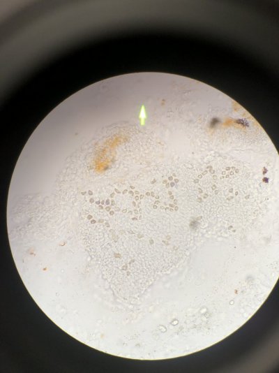
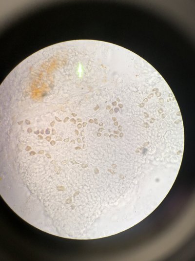
This lesion was first noted in first week of Jan. We performed several wet mounts and looked under microscope and had no findings. The wound was open and fleshy, so we decided to treat topically with neosporin once a day. We stopped treatment at day 12 because her wound was almost completely healed and she had stopped eating. We were unsure if this was because of separation from the male or from the stress of us treating her once a day (which is another reason why we stopped treatment). After that, she's been eating regularly and the wound was gone.
Until Feb 18th when the wound reappeared. In Jan, we assumed it was a wound from fighting. Although it is in an unusual spot for a fighting wound, the consistency in the tank (no new tank mates, consistent water quality with weekly water changes) we concluded fighting as the main culprit. Now we do not know. Performed more wet mounts, no findings. This time I did not treat with neosporin to see what happens, and the lesion went away on its own.
Like I stated, both clowns have been in this tank for at least 5 yrs. They have a reputation for fighting, so they've had injuries and lesions before, but this is different. The clowns also laid eggs regularly. Separating them has shown that this particular lesion is not from fighting, and honestly the smaller male is thriving having an anemone all to himself in his new tank. He has significantly improved (not just from his injuries, but color is more vibrant and he's very active). No concerns have been noted in the other fish in tank that he was moved to. I cannot say the same for the female, her color is not as vibrant, but she is at least eating regularly.
Theories include:
Parameters from yesterday:
ph 8.0, ammonia 0, nitrites 0, nitrates 0, phos 0, Ca 500+, Alk 8, salinity 1.026 (we have no ATO so I manually top off when I feed), temp 79F
Please note this is a school tank and we are using the API kits.
Weekly 10 gal water changes with Instant ocean reef crystals.
Attached are photos from different days/times so you can see the progression of the lesion. (I know they are blurry, I apologize in advance).
What do you guys think? Thank you for your time! Will post any updates as they happen :)


I am a tank caretaker at our college and we have this interesting case I wanted to share in case anyone recognizes it. This female clown is in a 30gal SW tank with a purple dottyback. Other inhabitants are really large toadstool, CUC (urchin, green emerald crabs, hermits, snails, etc), some hammer coral, and lots of manjano anemones (I know! they don't look that bad though). The male clown was removed because of continuous fighting. Both clowns and the dottyback have been in this tank for over 5yrs (that we know of, every year a new student takes care of the tank). The clowns were separated in Jan 2021 because the male was continuously beat up, the cranial half of the dorsal fin was actually devitalized and sloughed off. He is with a foster and is doing great. However, the female continuous to present with this recurring lesion near her R pectoral fin.
This lesion was first noted in first week of Jan. We performed several wet mounts and looked under microscope and had no findings. The wound was open and fleshy, so we decided to treat topically with neosporin once a day. We stopped treatment at day 12 because her wound was almost completely healed and she had stopped eating. We were unsure if this was because of separation from the male or from the stress of us treating her once a day (which is another reason why we stopped treatment). After that, she's been eating regularly and the wound was gone.
Until Feb 18th when the wound reappeared. In Jan, we assumed it was a wound from fighting. Although it is in an unusual spot for a fighting wound, the consistency in the tank (no new tank mates, consistent water quality with weekly water changes) we concluded fighting as the main culprit. Now we do not know. Performed more wet mounts, no findings. This time I did not treat with neosporin to see what happens, and the lesion went away on its own.
Like I stated, both clowns have been in this tank for at least 5 yrs. They have a reputation for fighting, so they've had injuries and lesions before, but this is different. The clowns also laid eggs regularly. Separating them has shown that this particular lesion is not from fighting, and honestly the smaller male is thriving having an anemone all to himself in his new tank. He has significantly improved (not just from his injuries, but color is more vibrant and he's very active). No concerns have been noted in the other fish in tank that he was moved to. I cannot say the same for the female, her color is not as vibrant, but she is at least eating regularly.
Theories include:
- This is some sort of Mycobacteriosis. Noga's book on Fish Diseases states that the bacteria can survive for 2 years in the environment. Non-healing, shallow to deep skin ulcers is a clinical sign as well. However, no granulomas have been noted on wet mount.
- Another theory is that she is self mutilating herself by trying to get into a spot in the rocks that she may have fit when she was much smaller. Thus she gets the same injury. However, I have been caring for this tank since Aug 2020 and haven't seen it before.
- Another theory is that maybe the dottyback is getting aggressive and fighting with her too. The dottyback was in poor condition when we took over tank care, we changed over to frozen food, and he's gone from almost white to the a purple color. He had no lesions, he was just white. He's always been kind of skiddish.
- Don't think its Brooklynella because that tends to be a very acute disease that causes mortality quickly, and we have not seen it on any wet mounts we have performed.
Parameters from yesterday:
ph 8.0, ammonia 0, nitrites 0, nitrates 0, phos 0, Ca 500+, Alk 8, salinity 1.026 (we have no ATO so I manually top off when I feed), temp 79F
Please note this is a school tank and we are using the API kits.
Weekly 10 gal water changes with Instant ocean reef crystals.
Attached are photos from different days/times so you can see the progression of the lesion. (I know they are blurry, I apologize in advance).
What do you guys think? Thank you for your time! Will post any updates as they happen :)
Last edited: