Dynos or Cyano?

soccersw

New Member
View Badges
Joined
Jul 17, 2026
Messages
8
Reaction score
0
Rating - 0%
0   0   0
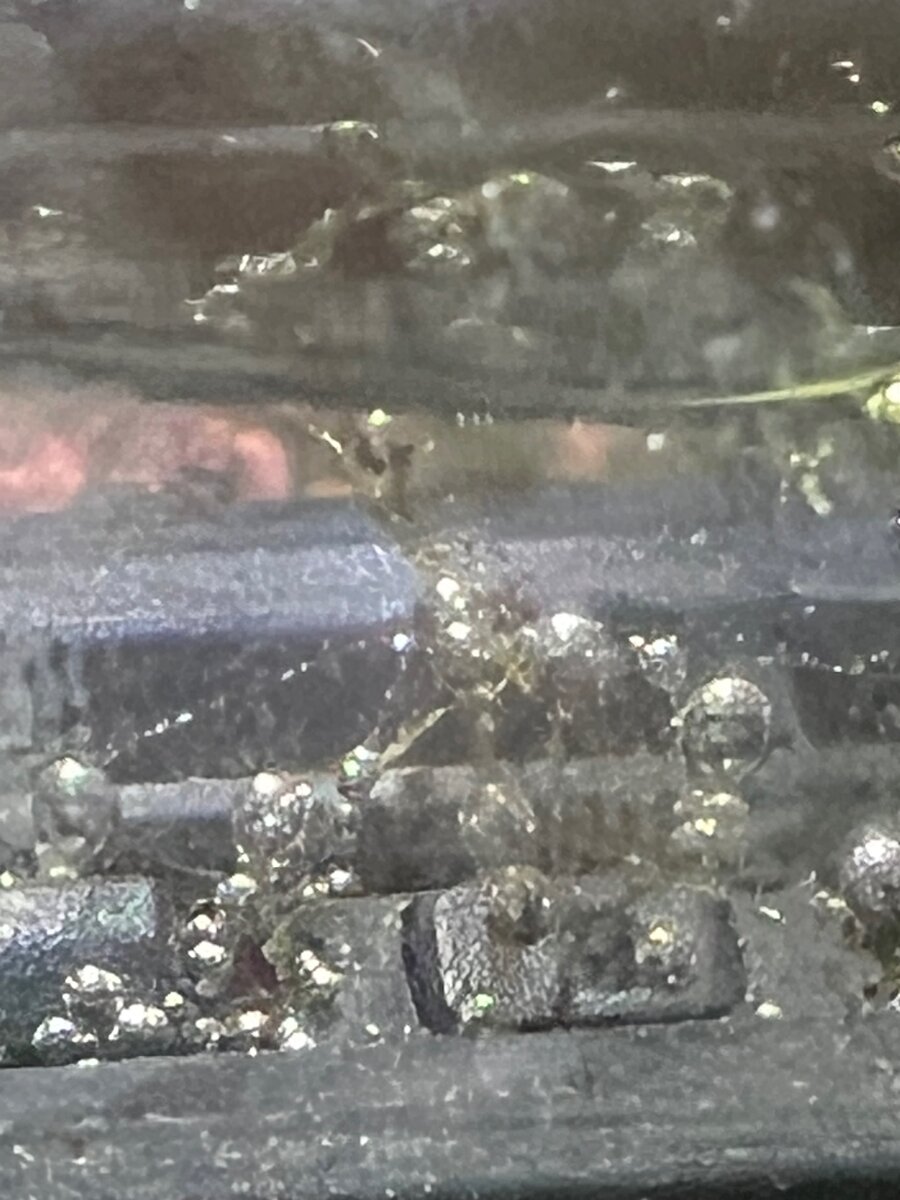
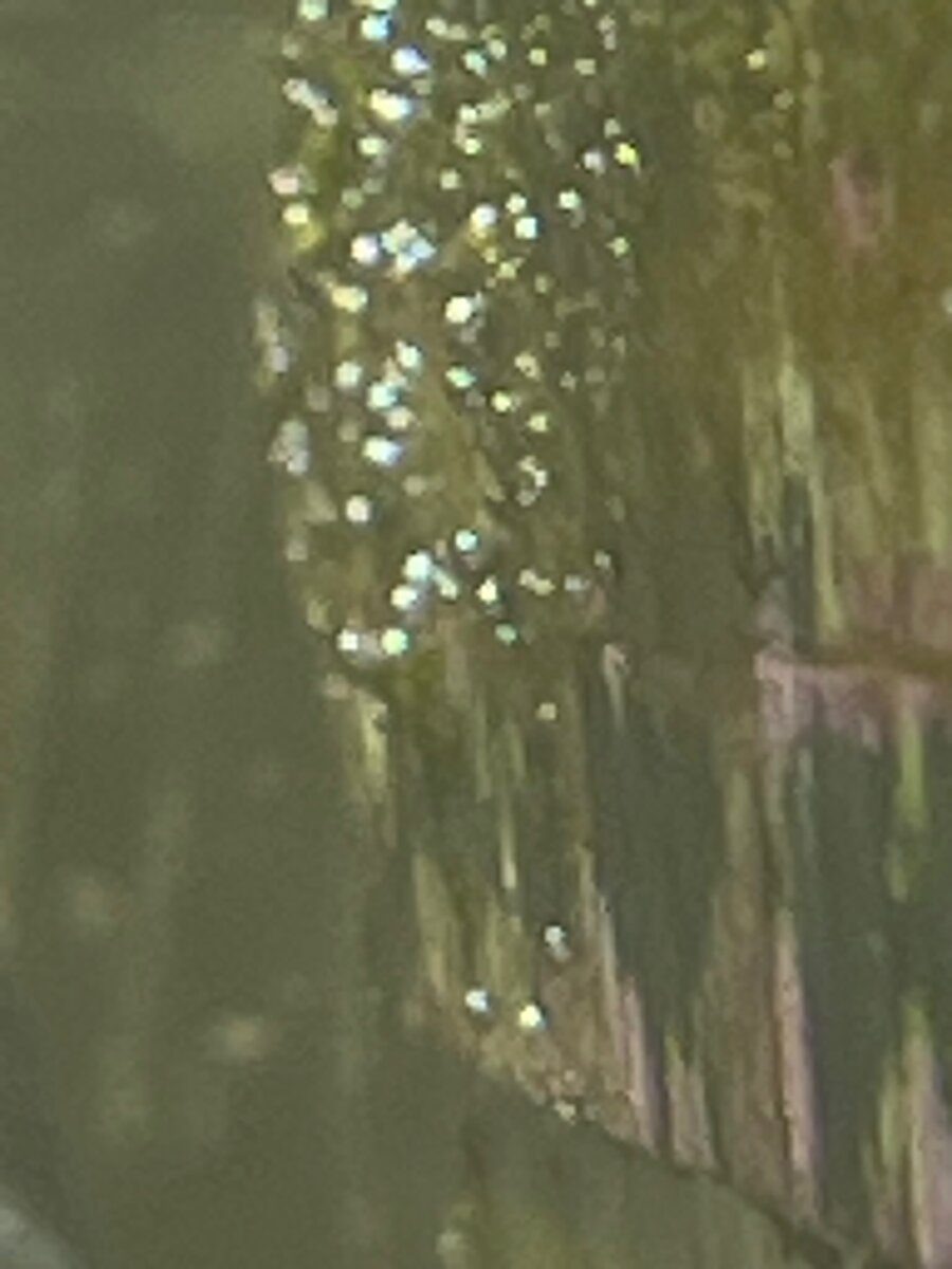
I have been fighting this for a few months. The stuff in the sand seems to clump and is very hard to vacuum because it is as heavy as the sand. I have used Red slime remover, with not much help. My nutrients are Nitrates 12 Phosphates .06 Salinity 35 Alkalinity 9.6. I am dosing Red Sea four part and have been happy with tank for years. Coral do not seem to be affected much. Tangs do not seem to eat it. My clean up crew do not seem to live very long. My sand sifting starfish looked like something was biting it. Can someone help me please?
IMG_A5557691-1F8C-4A3D-A8E3-9DC7F7C87F9F.jpeg
IMG_5546.jpeg
IMG_5548.jpeg
 
Could be another Euglenida case...
A compound microscope would help with the identification, but this would explain why your CUC dies.
Thank you for your information. I ordered a microscope today. Time to stop guessing and get some empirical evidence!
Just to be sure: It's not a digital one is it? It's a classical compound microscope like you may have had in school, right?

I'm asking because digital microscopes often don't have the required magnification or resolution capabilities.

You won't regret the purchase, microscopy in combination with a reef tank is pretty fun: https://www.reef2reef.com/threads/microscopy-thread.1138845/
 
Upvote 0
Just to be sure: It's not a digital one is it? It's a classical compound microscope like you may have had in school, right?

I'm asking because digital microscopes often don't have the required magnification or resolution capabilities.

You won't regret the purchase, microscopy in combination with a reef tank is pretty fun: https://www.reef2reef.com/threads/microscopy-thread.1138845/
Just a cheaper 2000 x classic compound microscope. Should be plenty strong for this application. I will keep the electron microscope for later. Lol
 
Upvote 0
Just a cheaper 2000 x classic compound microscope. Should be plenty strong for this application. I will keep the electron microscope for later. Lol
There should be a 20x and a 40x objective together with 10x eye piece. These will probably work best for the identification. It doesn't matter much that it's cheap, it will probably do just fine 😊
Tag me once you have some images, I'm curious what you will find! You are also welcome to post your findings in my microscopy thread if you want.

Side note:
2000x is just marketing.
You won't be able to go much further than 1000x with light microscopy. Yes, if you have a 100x objective and a 20x eye piece you theoretically should end up with 2000x, but the image will be too dark to be useful and the resolution won't increase beyond 1000x making it like a "digital zoom" which doesn't provide more information. The wavelength of visible light simply won't allow for higher resolution.
 
Last edited:
Upvote 0
Could be another Euglenida case...
A compound microscope would help with the identification, but this would explain why your CUC dies.
I posted my experience in the thread above where I outline my experience dealing with this kind of green toxic growth on the sand bed for the last six weeks. I am on day 3 of removing the patches of sand with green growth and top dressing with ocean live sand from Tampa Saltwater. So far the sand looks immaculate with no signs of the green algae. Notably, during the first there were small patches of sand in remote areas of the reef scape. I mixed those areas up so they came into more contact with the TBS sand. It is day 3 now and no sign of algae regrowth. The sand is basically complex clear!
 
Upvote 0
There should be a 20x and a 40x objective together with 10x eye piece. These will probably work best for the identification. It doesn't matter much that it's cheap, it will probably do just fine 😊
Tag me once you have some images, I'm curious what you will find! You are also welcome to post your findings in my microscopy thread if you want.

Side note:
2000x is just marketing.
You won't be able to go much further than 1000x with light microscopy. Yes, if you have a 100x objective and a 20x eye piece you theoretically should end up with 2000x, but the image will be too dark to be useful and the resolution won't increase beyond 1000x making it like a "digital zoom" which doesn't provide more information. The wavelength of visible light simply won't allow for higher resolution.
Thank you for all your information. I didn't get a microscope with imaging. I will share the information, if I can match what I see to a known strand or algae.
 
Upvote 0
I posted my experience in the thread above where I outline my experience dealing with this kind of green toxic growth on the sand bed for the last six weeks. I am on day 3 of removing the patches of sand with green growth and top dressing with ocean live sand from Tampa Saltwater. So far the sand looks immaculate with no signs of the green algae. Notably, during the first there were small patches of sand in remote areas of the reef scape. I mixed those areas up so they came into more contact with the TBS sand. It is day 3 now and no sign of algae regrowth. The sand is basically complex clear!
I will keep an eye out for how your's is going. I guess you have taken away the light and provided beneficial bacteria to the system. Sounds like a good idea. Do I have the general Idea of what you are doing?
 
Upvote 0
There should be a 20x and a 40x objective together with 10x eye piece. These will probably work best for the identification. It doesn't matter much that it's cheap, it will probably do just fine 😊
Tag me once you have some images, I'm curious what you will find! You are also welcome to post your findings in my microscopy thread if you want.

Side note:
2000x is just marketing.
You won't be able to go much further than 1000x with light microscopy. Yes, if you have a 100x objective and a 20x eye piece you theoretically should end up with 2000x, but the image will be too dark to be useful and the resolution won't increase beyond 1000x making it like a "digital zoom" which doesn't provide more information. The wavelength of visible light simply won't allow for higher resolution.

IMG_5563.jpeg
 
Upvote 0
There should be a 20x and a 40x objective together with 10x eye piece. These will probably work best for the identification. It doesn't matter much that it's cheap, it will probably do just fine 😊
Tag me once you have some images, I'm curious what you will find! You are also welcome to post your findings in my microscopy thread if you want.

Side note:
2000x is just marketing.
You won't be able to go much further than 1000x with light microscopy. Yes, if you have a 100x objective and a 20x eye piece you theoretically should end up with 2000x, but the image will be too dark to be useful and the resolution won't increase beyond 1000x making it like a "digital zoom" which doesn't provide more information. The wavelength of visible light simply won't allow for higher resolution.

IMG_5563.jpeg
Ostreopsis sp. (Dinoflagellates)

And detritus... Weird color for Ostreopsis though. Usually they are brown but without the microscope it looks green. Have you tried getting a single sand grain under the scope?
 
Last edited:
Upvote 0
Ostreopsis sp. (Dinoflagellates)

And detritus... Weird color for Ostreopsis though. Usually they are brown but without the microscope it looks green. Have you tried getting a single sand grain under the scope?

 
Upvote 0
Ostreopsis sp. (Dinoflagellates)

And detritus... Weird color for Ostreopsis though. Usually they are brown but without the microscope it looks green. Have you tried getting a single sand grain under the scope?
According to AI it implied it was Amphidinium, because of the smooth, glide, swimming pattern. I had uploaded several still images to AI also.
 
Upvote 0
According to AI it implied it was Amphidinium, because of the smooth, glide, swimming pattern. I had uploaded several still images to AI also.
I'm sure AI is wrong in this case. It's Ostreopsis. It has a pointy end and not a "beak" or "nose" like Amphidinium has. It also tries to move in circles around a point in the distance which the tip points towards, but in your video the cells struggle to do this as they crash into each other 😅

1785093868542.png

(Ostreopsis sp. from my initial microscopy thread post)

1785093991180.png
1785093997866.png

(Amphidinium cells from post #3 in my microscopy thread)​
 
Upvote 0

TOP 10 Trending Threads

ARE YOU READY TO CONFESS TO CRAZIEST, DUMBEST, FUNNIEST THING YOU’VE EVER DONE IN REEFING?

  • Yeah, I'll confess! (Share your story in the comments!)

    Votes: 38 54.3%
  • Nah, I'll keep mine a secret...(Don't be like that, share with the class!)

    Votes: 32 45.7%
Back
Top
Home
Post thread…
Market
What's new